- Home
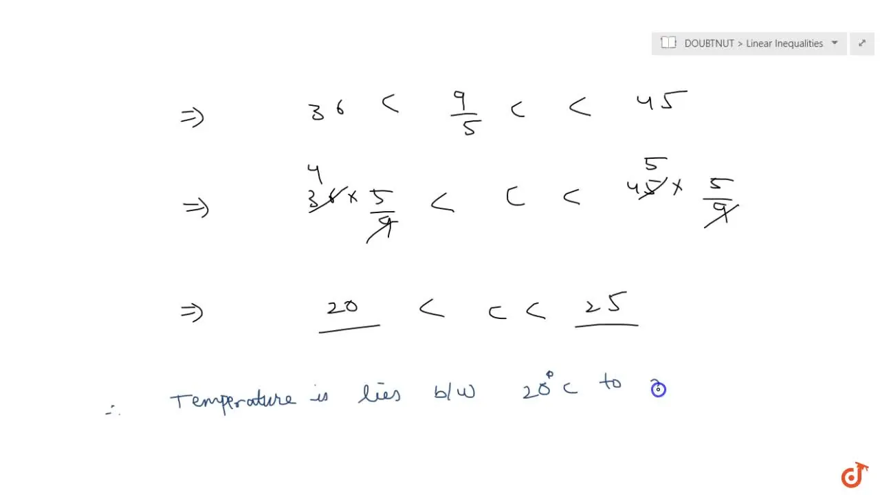
- 36 c in f
- A) Preoperative intraoral periapical (IOPA) radiograph of 36. B) Post operative (IOPA) radiograph of 36. C) 1 month follow up IOPA radiograph of 36. D) 6 months follow up IOPA radiograph of
A) Preoperative intraoral periapical (IOPA) radiograph of 36. B) Post operative (IOPA) radiograph of 36. C) 1 month follow up IOPA radiograph of 36. D) 6 months follow up IOPA radiograph of
4.7 (653) · $ 5.99 · In stock

A) Preoperative intraoral periapical (IOPA) radiograph of 36. B) Post operative (IOPA) radiograph of 36. C) 1 month follow up IOPA radiograph of 36. D) 6 months follow up IOPA radiograph of 36. E) 1 year follow up IOPA radiograph of 36. - IP Indian J Conserv Endod - clinical and preclinical conservative /restorative de

Nonsurgical Management of Periapical Lesion: A Case Series

JaypeeDigital

Treatment of Deeply Carious Vital Primary Molars by Complete

Treatment of Deeply Carious Vital Primary Molars by Complete Caries Removal Using Three Different Bioactive Materials: A Pilot Study

Oral Radiology - Common Types Of Intraoral Radiograph

IOPA at three months postoperative. Distinct borders between graft
Cureus, Mandibular Alveolar Ridge Split With Simultaneous Implant Placement: A Case Report

PDF) Direct pulp capping with bioactive materials – A case series

IOPA radiograph (post-op) of after 6 month follow-up showing almost

a) Preoperative IOPA radiograph of tooth #36. (b) Intraoral image

Preoperative and postoperative intraoral radiographs a: Preoperative

A Preoperative intraoral peri-apical (IOPA) radiograph of lower left

Radiograph sem
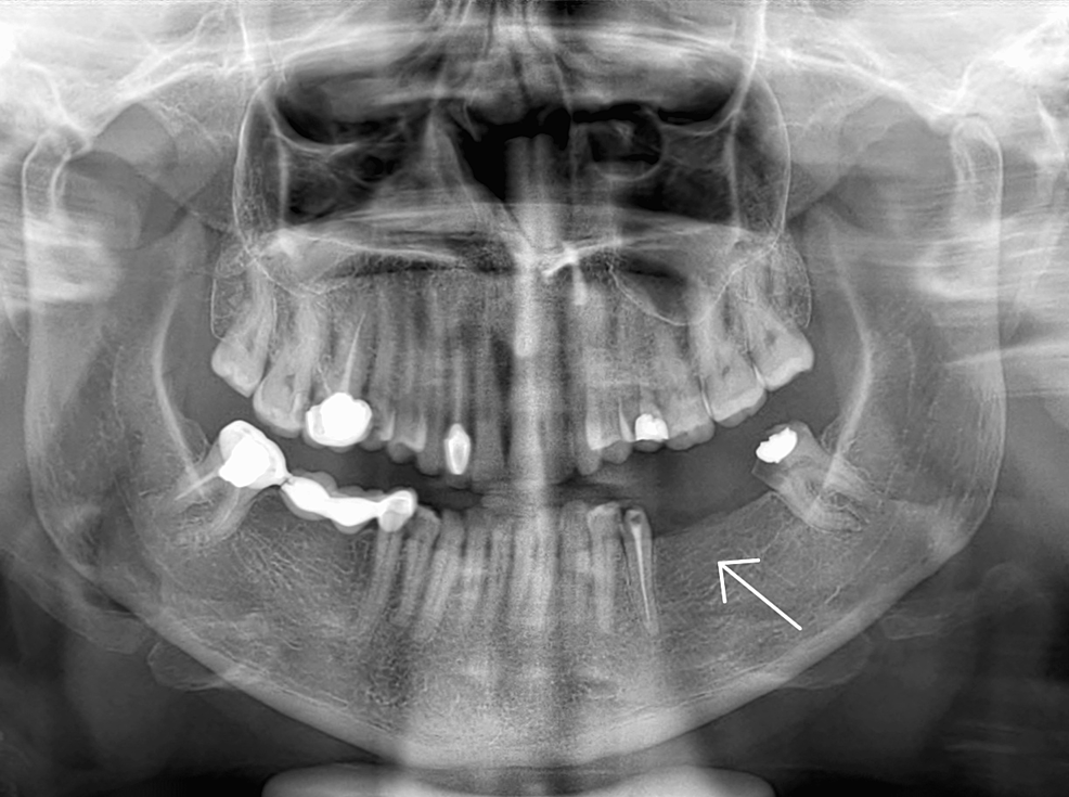
A) Intraoral periapical (IOPA) radiograph of blocked mandibular right


:quality(70)/cloudfront-us-east-1.images.arcpublishing.com/metroworldnews/H6HYWP7GRFECTB2Q6ZTNVK7UX4.JPG)
---ThoughtCo.webp)