Results of a 32-year-old female nulliparous patient The body mass
4.8 (227) · $ 17.00 · In stock

Download scientific diagram | Results of a 32-year-old female nulliparous patient The body mass index of the patient was 20.4 kg/m 2. The oncologic breast surgeon performed skin-sparing mastectomy. (A) Preoperative view. (B) Postoperative view at 6 months of follow-up. (C) Postoperative view at 12 months after nipple reconstruction. from publication: Weight analysis of mastectomy specimens and abdominal flaps used for breast reconstruction in Koreans | Background: Slim patients or those with large breasts may be ineligible for breast reconstruction with an abdominal flap, as the volume of the flap may be insufficient. This study aimed to establish that abdominal tissue-based breast reconstruction can be well suited for | Breast Reconstruction, Mammaplasty and Free Tissue Flaps | ResearchGate, the professional network for scientists.
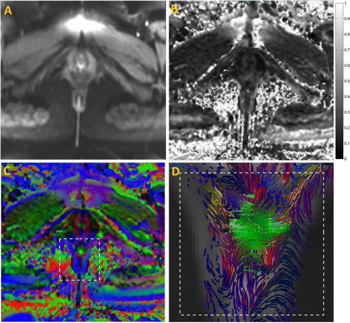
Connectivity of the Superficial Muscles of the Human Perineum: A Diffusion Tensor Imaging-Based Global Tractography Study
Fecal Incontinence Treatment & Management: Medical Therapy, Surgical Therapy, Preoperative Details
Cancers, Free Full-Text

Fecal incontinence in nonpregnant nulliparous women aged 25 to 64 years-a randomly selected national cohort prevalence study - ScienceDirect

Maternal body mass index in pregnancy and mental disorders in adult offspring: a record linkage study in Aberdeen, Scotland
Incidence and risk factors for Preeclampsia in a cohort of healthy nulliparous pregnant women: a nested case-control study
Nutrients, Free Full-Text

Demographic data for the whole sample

Prediction of pre-eclampsia in nulliparous women using routinely collected maternal characteristics: a model development and validation study, BMC Pregnancy and Childbirth

Evidence on: Pregnancy at Age 35 and Older - Evidence Based Birth®
NBME OBGYN Form 4 – Step Prep

Jin Sup Eom's research works University of Ulsan, Ulsan (UOU) and other places
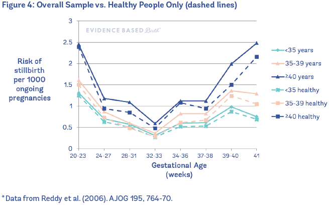
Evidence on: Pregnancy at Age 35 and Older - Evidence Based Birth®

Hyung Hwa Jeong's research works University of Ulsan, Ulsan (UOU) and other places

Combination of tomographic ultrasound imaging and three-dimensional magnetic resonance imaging-based model to diagnose postpartum levator avulsion